Please activate JavaScript!
Please install Adobe Flash Player, click here for download
ePaper created 2012-07-20, 16:26:01 | version 1.24.0
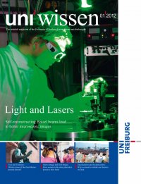
Less scattering: Alexander Rohrbach’s self-recon- structing laser beams remain stable in the center longer when they penetrate an object. In order to obtain even better microscopic images, he only uses the central main beam of the laser (small picture at upper left, diagram on page 22). nate objects like cancer cell clusters, insulated skin, or animal embryos, which scatter the light too strongly due to their size. The laser beam loses its original concentrated form to scattering due to a multitude of tiny particles or is deflected, making it almost impossible for the microscope to illuminate anything at all on the far side of the object. Rohrbach, a trained physicist, has been investigating biological systems for many years but has not always had success in extracting all of the desired information from them. For example, it is not yet clear when and how various forms of energy are built up inside the cell. “We will need to develop new microscopy techniques, approaches, and analyses to find the answers to these questions,” explains Rohrbach – and these prospects excite his passion for research again and again. Like several earlier research groups, the Frei burg professor has taken up the over-a-century-old idea of ultramicroscopy. Ultramicroscopy, or light sheet microscopy as it is called today, only illumi nates the objects at a particular level: the level to which the focus of the microscope’s objective lens is set. This is made possible by a light sheet,